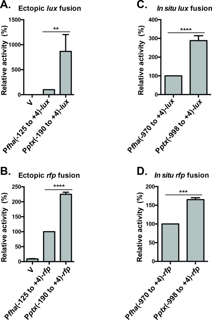
Fig 7. Comparing the relative strengths of Pfha and Pptx by transcriptional fusion.
Promoter-lux (A & C) and promoter-rfp (B & D) transcriptional fusions were constructed and integrated into the BP536 chromosome. Fusions to the lux operon used either pSS3967, to integrate at an ectopic location (A), or pSS4162, to integrate in situ (C). Similarly, transcriptional fusions to rfp used either pQC2241 for ectopic insertion (B) or pQC2319 for in situ insertion (D). For the two ectopic constructs “V” indicates insertion of the vector alone. This control is not possible for the in situ insertions. The extent of the promoter sequences cloned in each construct are provided as nucleotide coordinates relative to the transcriptional start. B. pertussis strains carrying these constructs were grown on BG agar at 37°C for 2 days and analyzed for and luciferase and RFP activity as described in Materials and Methods. In each panel activity is reported relative to the Pfha-promoter fusion and the results of at least four assays were used in the calculation of standard deviations and statistical analysis by an unpaired two-tailed t test between two samples. Statistical symbols are: **, P ≤ 0.01; ***, P ≤ 0.001; ****, P ≤ 0.0001.

