. 2020 Apr 30;9:e56656. doi: 10.7554/eLife.56656

Figure 2. Reconstitution of Skap2-/- mice with WT bone marrow hematopoietic stem cells confers protection against K. pneumoniae.

(A) Schematic used to generate bone marrow chimeras in (B–D, F). (B–F) Mice were infected with 5 × 10³ cfu K. pneumoniae (red asterisk); 24 hpi mice were sacrificed and lungs harvested. (E–F) WT and Skap2-/- mice were injected intraperitoneally with 50 μg of α-Ly6G (1A8) or 20 μg of α-CCR2 (MC-21) to deplete neutrophils and iMOs, respectively, or PBS 16 hr prior to infection. Bacterial burden (B, E, F) and percent live neutrophils (CD11b+ Ly6Ghi) (C), or inflammatory monocytes (CD11b+ Ly6Chi) (D) from K. pneumoniae-infected lungs. Data are compiled from 2 to 4 independent experiments using groups of 2–3 mice/genotype/experiment. Each dot represents a mouse, bars are geometric means (B, E–F) or means (C–D). Statistics were assessed using one-way ANOVA with (C–D) Sidak’s post-test, or (B, E–F) Tukey’s post-test. (D) Percent of iMOs were compiled, and comparison between WT and Skap2-/- irradiated recipient disregarding the donor BM were assessed by two-tailed unpaired Student’s t test.

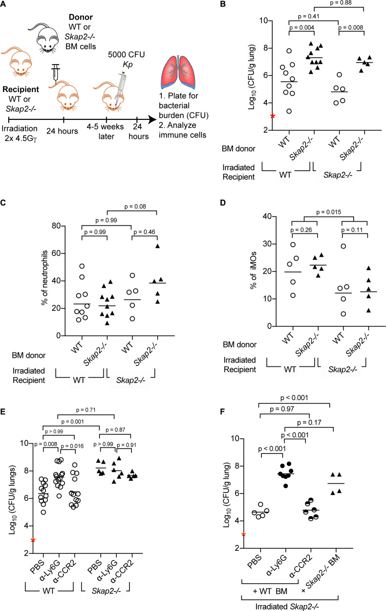
Figure 2.

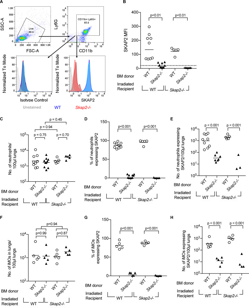
Figure 2—figure supplement 1. Transplantation efficiency of bone marrow reconstitution into WT and Skap2-/- recipients.

Figure 2—figure supplement 1.

(A) Schematic for gating strategy of flow cytometry data in bone marrow chimeras. Blocks and arrows indicate the cell population used for next gating step. (B–H) Neutrophil (CD11b+ Ly6Ghi) (C–E), or inflammatory monocytes (CD11b+ Ly6Chi) (F–H) from K. pneumoniae-infected lungs were unstained or intracellularly stained with SKAP2 antibody or isotype control. (B) SKAP2 expression in CD11b+ cells shown as mean fluorescent intensity (MFI). Dotted line indicates average level from cells intracellularly stained with isotype control. Data are compiled from 2 to 4 independent experiments using 2–3 mice/genotype/experiment. Each dot represents a mouse, bar represents means. Statistical significance was assessed using one-way ANOVA with Sidak’s post-test.
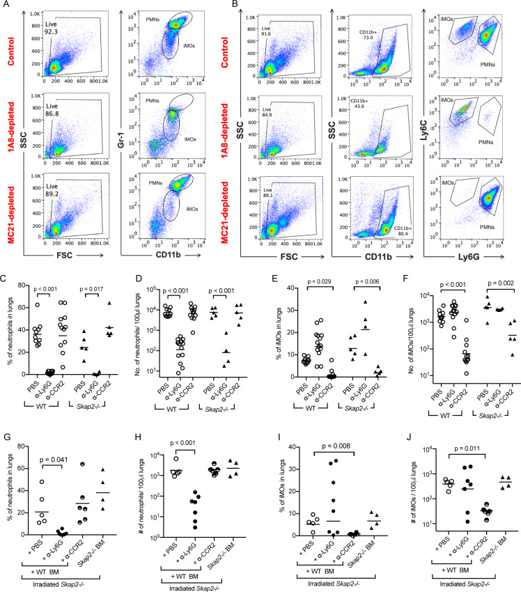
Figure 2—figure supplement 2. Evaluation of immune cell populations in depletion studies in WT and Skap2-/- mice.

Figure 2—figure supplement 2.

Analysis of inflammatory monocytes (CD11b+ Gr1lo or CD11b+ Ly6Chi Ly6Glo) and neutrophils (CD11b+ Gr1hi or CD11b+ Ly6Cint Ly6Ghi) from non-depleted (PBS), α-Ly6G (clone 1A8)-depleted, or MC-21-depleted (α-CCR2) K. pneumoniae-infected lungs. (A–B) Example of flow cytometry analysis for depletion experiments. (C–F) Quantification of neutrophils (CD11b+ Gr1hi) (C–D), or inflammatory monocytes (CD11b+ Ly6Chi Ly6Glo) (E–F) from α-Ly6G, α-CCR2, or PBS-treated, and K. pneumoniae-infected WT or Skap2-/- mice based on flow cytometry analysis. (G–J) Quantification of neutrophils (G–H), or inflammatory monocytes (I–J) from α-Ly6G, α-CCR2, or PBS-treated, K. pneumoniae-infected irradiated Skap2-/- mice based on flow cytometry analysis. Data are compiled from 3 to 4 independent experiments with 2–3 mice/genotype/experiment. Each dot represents a mouse, bars represent geometric means. (D, F, H, J) were log-transformed. Significance was assessed using one-way ANOVA with Tukey’s post-test.