. 2020 Apr 30;9:e56656. doi: 10.7554/eLife.56656

Figure 3. BM and DIV neutrophils require SKAP2 for integrin-activated ROS production, but not for FcγR.

(A–F) Extracellular respiratory burst of BM and DIV neutrophils. WT or Skap2-/- neutrophils were plated on a poly-RGD-coated surface (A–C), or IgG immune complex (IC)-coated surface (D–F), and superoxide production was measured by cytochrome C reduction. Unstimulated (unstim) cells were plated onto 10% FBS/PBS or stimulated with 100 nM PMA. (C, F) Total concentration of superoxide produced after 60 min. (G–J) Expression of surface receptors on DIV neutrophils of CD11b (G–H) or activating CD16 Fcγ receptor (I–J). (G–J); Blue shaded areas and bars represent WT, and red, Skap2-/-. (A–B, D–E) represent the mean ± SD of one experiment in technical triplicate assessed using two-way ANOVA with Tukey’s post-test. (C, F, H, J) represent the mean ± SEM of at least three independent experiments performed in at least technical duplicate (C, F) and were assessed using two-way ANOVA with Tukey’s post-test (C, F), or two-tailed unpaired Student’s t test (H, J).

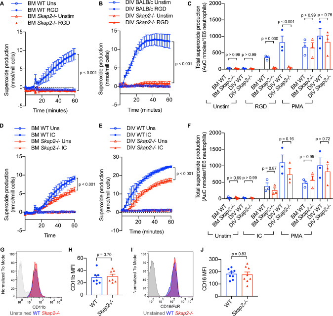
Figure 3.

Figure 3—figure supplement 1. Hoxb8-transformed GMP differentiate into neutrophils with similar morphology and surface markers to that of BM-derived neutrophils.

Figure 3—figure supplement 1.

(A) Schematic of MSCV-Hoxb8 transduction system to generate immortalized stem cell progenitors. The estrogen-binding domain (ERBD) of the estrogen receptor fused to Hoxb8 with an N-terminal Flag epitope tag allows conditional expression of Hoxb8 in the presence of estrogen. After 4 days of differentiation, cells are referred to as DIV neutrophils. (B) Nuclear morphology of BM-derived neutrophils isolated from WT or Skap2-/- mice, HoxB8 GMP, and DIV neutrophils with DAPI. Red arrows indicate polymorphonuclear nuclei. (C) Analysis of c-Kit and CD11b expression. (D) Analysis for Ly6G expression on live CD11b+ cells. (E–F) Viability of WT and Skap2-/- DIV neutrophils was assessed using (E) trypan blue exclusion test from neutrophils differentiated from Hoxb8 immortalized GMP from 2 different WT and Skap2-/- mice prior to functional studies, or (F) DIV neutrophils were plated into 10%FBS/PBS-coated wells, loaded with CellTiter Glo reagent, and chemiluminescence was detected for 30 min by plate reader. The quantification of ATP is shown as relative light units (RLU) from one experiment done in technical triplicate. (G–I) Quantification of (G) cKit+, (H) CD11b+, or (I) CD11b+ Ly6G+ cells from Hoxb8 GMP, day 4 DIV neutrophils (DIV), and BM neutrophils (BM). Results are compiled from 1 to 4 independent experiments with each dot representing one experiment and are shown as mean ± SEM. Significance was determined by one-way ANOVA with Sidak’s post-test.