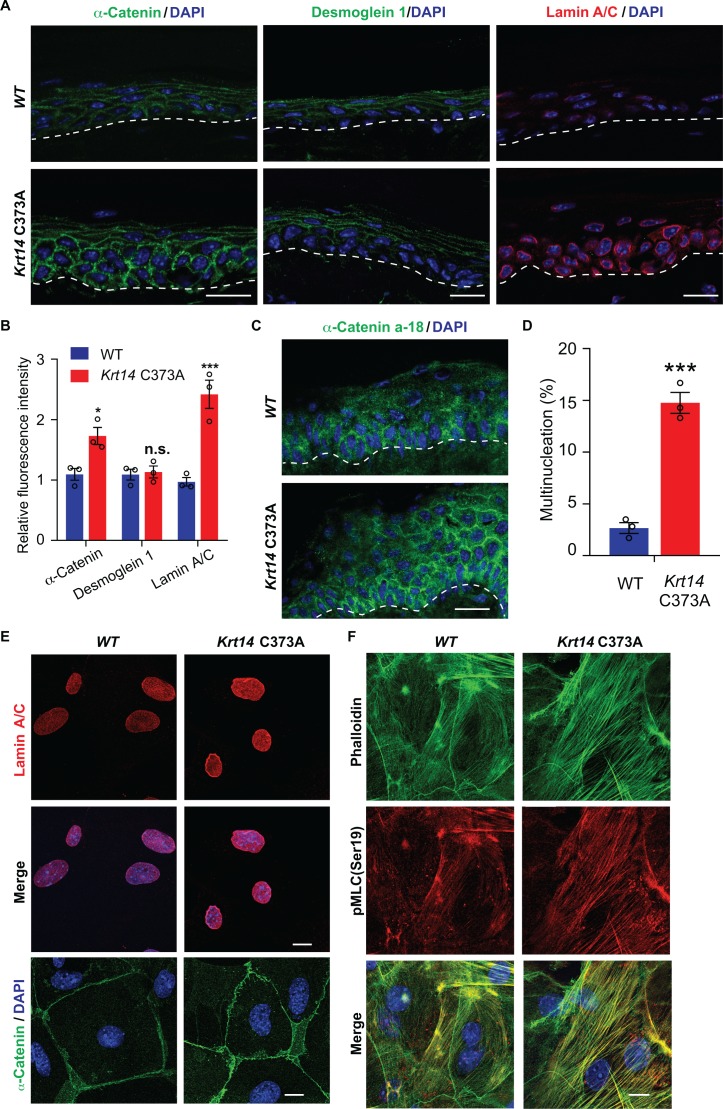
Figure 6. Altered mechanics in Krt14 C373A epidermis and keratinocytes in culture.
(A-C) Studies involving tail skin sections from young adult WT and Krt14 C373A mice. A. Indirect immuno-fluorescence microscopy for α-catenin, desmoglein one and lamin A/C. Dashed lines depict the dermo-epidermal interface. B. Quantification of relative fluorescence intensity, as shown in frame a, for WT and Krt14 C373A. N = 3 biological replicates. (C) Indirect immunofluorescence microscopy for the a-18 mechanosensitive epitope in α-catenin in tail skin sections from WT and Krt14 C373A mice (see A). D-F: Studies involving newborn skin keratinocytes in primary culture. (D) Percentage of cells with multinucleation in WT and Krt14 C373A keratinocytes cultured as described for frames a,c. N = 3 biological replicates (total of 100 cells counted each time per genotype). (E) Indirect immunofluorescence microscopy for lamin A/C (top and middle rows) and α-catenin (bottom row) in primary cultures of WT and Krt14 C373A newborn keratinocytes. (F) same as E, except that F-actin (via phalloidin) and Ser19-phosphorylated myosin light chain pMLC (Ser19) are stained. Nuclei are stained with DAPI in frames A, C, E and F. Scale bars, 20 µm. Data in B and F represent mean ± SEM. Student’s t test: *p<0.05; ***p<0.005; n.s., no statistical difference.

