. 2020 May 26;9:e55487. doi: 10.7554/eLife.55487

Figure 4. HIV preferentially infects endometrial Tem and Trm with phenotypic features of Th1, Th2, and Tfh cells.

The proportions of ETs that were Tem (CCR7-CD62L-), Tcm (CCR7+CD62L+), Th1 (Tbet+), Th2 (CRTh2+), Th17 (RORγt+), Tregs (CD127-CD25+), Tfh (PD1+CXCR5+), and Trm (CD69+) were compared among uninfected Tm cells (UI) and PRE cells. *p<0.05, **p<0.01, ***p<0.001 as assessed using the Student’s paired t test. n.s.: not significant.
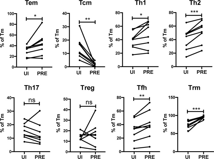
Figure 4.

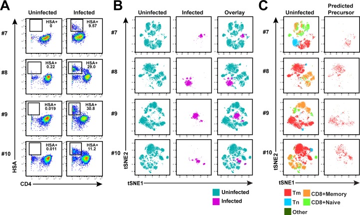
Figure 4—figure supplement 1. Memory CD4+ T cells from the endometrium of 4 additional donors are highly susceptible to productive infection by HIV.

Figure 4—figure supplement 1.

(A) ETs from 4 additional donors were mock-treated or inoculated with F4.HSA, and then monitored 3 days later by CyTOF for levels of productive infection. Left: Uninfected samples. Right: F4.HSA-exposed cells. Results are gated on live, singlet CD3+CD8- cells. (B) t-SNE plots corresponding to uninfected T cells (left), HIV-infected cells, and an overlay of the two plots (right) for the 4 donors shown in panel A. (C) t-SNE plots of uninfected T cells and the PRE cells from the 4 donors shown in panel A demonstrate that the dominant population of susceptible cells are Tm. Tm, Tn, memory CD8+ T cells, and naïve CD8+ T cells were identified by manual gating and colored as indicated.
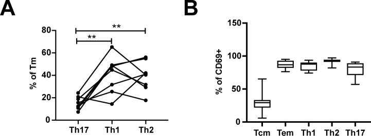
Figure 4—figure supplement 2. Frequencies of differentiated subsets expressing the Trm marker CD69.

Figure 4—figure supplement 2.

(A) Proportions of Tm that were Th17 (RORγt+), Th1 (Tbet+), or Th2 (CRTh2+) cells. **p<0.01 as assessed using the Student’s paired t test and adjusted for multiple testing using the Benjamini-Hochberg for FDR. (B) The proportions of Tcm (CCR7+CD62L+), Tem (CCR7-CD62L-), Th1 (Tbet+), Th2 (CRTh2+), and Th17 (RORγt+) cells that expressed the Trm marker CD69.