Figure 6.
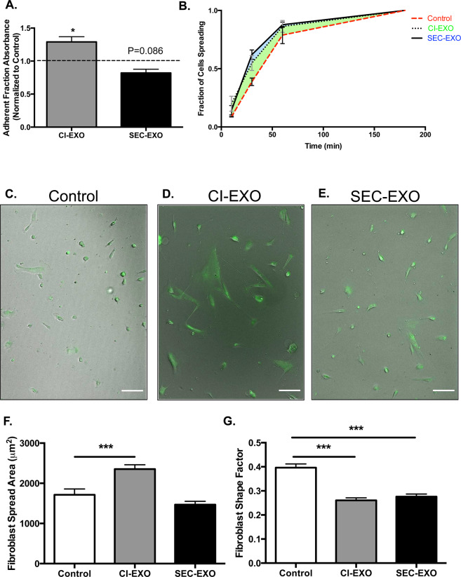
Exosome Treatment Regulates CAF Detachment and Spreading Profiles. CAFs were seeded overnight on FN-coated surfaces. Cells were treated with or without exosomes for 24 hours and detached using a centrifugal force-based assay. (A) Adherent fractions of fibroblasts treated with either population of exosomes were normalized to control groups. The dashed line indicates control group. Statistics calculated using Kruskal-Wallis test and were reported as mean +/− SEM. (B) To determine the fraction of cells spreading, CAFs were treated with exosomes for 24 hours, detached, and adhered on FN coated surfaces. The number of cells spreading over total number of cells at each time point was reported. (C–E) 10X images of calcein-labeled fibroblasts for each condition were taken to analyze cell spread profiles after 12 hours. Scale bar: 100 μm. (F) Fibroblast spread area and (G) shape factor were measured at the 12-hour time point (N = 3). Statistics calculated using Student’s t-test and reported as values +/− SEM. *p < 0.05, ***p < 0.001.

