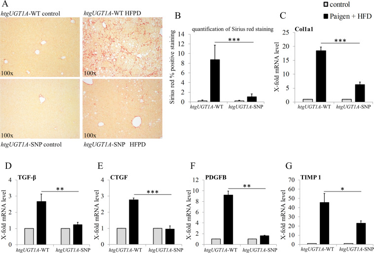
Figure 2.
Assessment of liver fibrosis in htgUGT1A-WT and SNP mice after high-fat Paigen diet (HFPD) treatment. (A) Representative hepatic sections of histological Sirius red staining and (B) computational quantification of Sirius red stained areas. Hepatic collagen deposition was significantly lower in htgUGT1A-SNP mice. (C–G) Gene expression levels of the profibrotic biomarkers collagen type 1 alpha 1 (Col1a1), transforming growth factor beta (TGF-β), connective tissue growth factor (CTGF), platelet-derived growth factor subunit B (PDGFB) and tissue inhibitor metalloprotease 1 (TIMP 1) as indicators for severity of liver fibrosis. Transcriptional activation of all depicted fibrosis marker genes was significantly lower in htgUGT1A-SNP mice. In case of TGF-β and CTGF, minor transcriptional inductions were detected in HFPD-treated htgUGT1A-SNP mice. Graphs are expressed as means ± standard deviation. *p < 0.05; **p < 0.01; ***p < 0.001.

