Figure 3.

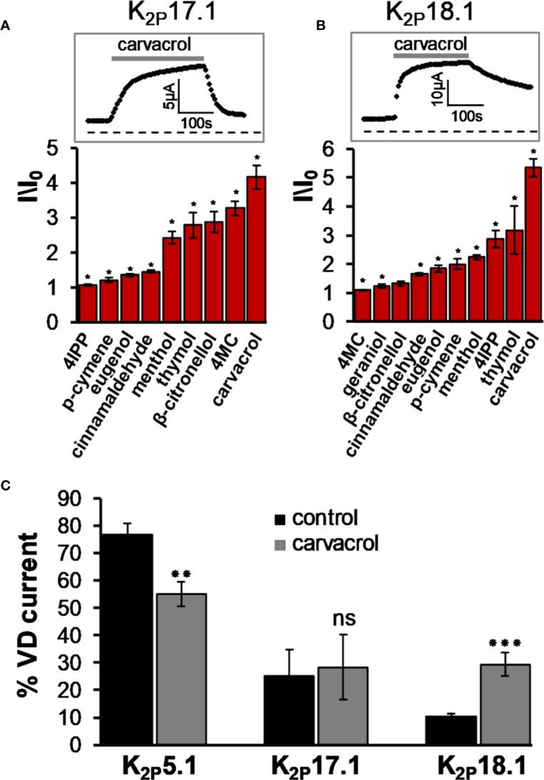
K2P17.1 and K2P18.1 are activated by monoterpenes. (A), (B) Activation of K2P17.1 (A) and K2P18.1 (B) by monoterpenes. Oocyte membrane potential was held at −80 mV and pulsed to +25 mV for 75 ms with 5 s interpulse intervals. All MTs were applied at the concentration of 0.3 mM, and currents were measured 4 min after application of the indicated monoterpene (mean ± S.E., n = 5–10). Insets: currents of representative oocytes expressing K2P17.1 (A) and K2P18.1 (B) during application of 0.3 mM carvacrol. (C) Fraction of voltage-dependent (VD) current (in %) under control conditions and after application of 0.3 mM carvacrol for three channel types, as indicated. Oocytes were held at −80mV, and currents were measured at 30 mV. A fit of the results to an exponential decay slope was used to identify the initial current (mean ± S.E., n = 6–9). *p ≤ 0.05, **p ≤ 0.01, ***p ≤ 0.001, ns, not significant.
