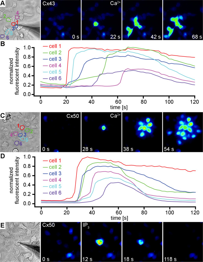
Figure 4.
Ca2+ and IP3 permeation in monolayer cultures of Cx43 and Cx50 expressing cells. After recording background fluorescence for ~20 seconds, 2 mM Ca2+ was released into a single cell in a cluster of Cx43 expressing cells loaded with Fluo-8 (A). Fluorescence rapidly increased in the patched cell, followed by an increase in 5 more distal cells (B) within 60 seconds. Similar (C–D) results were obtained when 2 mM Ca2+ was introduced into the cytoplasm of a single cell within a cluster of Cx50 expressing cells. In contrast, delivery of 500 µM IP3 into the cytoplasm of a single cell within a cluster of Cx50 expressing cells (E) resulted in a rapid peak of fluorescent intensity in the injected cell, with no evidence of IP3 permeation to any of the adjacent cells.

