. 2020 May 20;45(2):95–104. doi: 10.1584/jpestics.D20-001

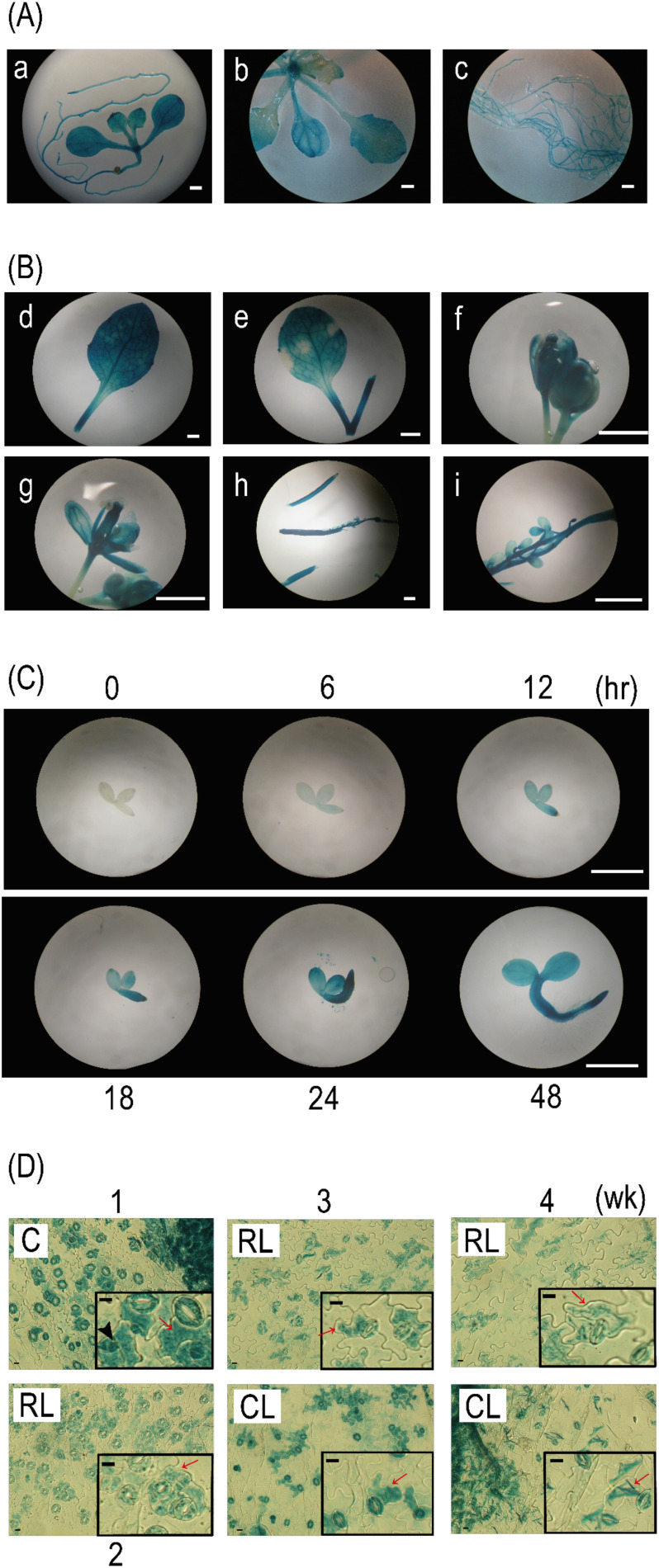
Fig. 5. BEH4::GUS plants were subjected to histochemical GUS staining at different stages: seedlings in an early growth stage (A, 1 wk and 2 wk), several organ parts excised from adult plants in a late growth stage (B, 4 wk or later), and seedlings in a very early stage (C, 0–48 hr). Leaf epidermal tissues (D) that were peeled off from the cotyledon (C, 1 wk), rosette leaf (RL, 2–4 wk), and cauline leaf (CL, 3 wk and 4 wk) of BEH4::GUS plants were subjected to GUS staining. The lowercase letters in A and B indicate a 1-wk-old whole seedling (a); above-ground organs (b) and roots (c) of 2-wk-old seedlings; a 4-wk-old rosette leaf (d); a 4-wk-old cauline leaf (e); a 5-wk-old flower bud (f); an open flower (g); an immature silique (h); and a developing seed in a silique (i). Insets in Fig. 5D present magnified images of the epidermal tissues. White bars and black bars represent 1 mm and 10 µm, respectively. Red arrows and black arrowheads indicate pavement cells (PCs) located adjacently to guard cells (GCs) and smaller cells located away from GCs, respectively.

Fig. 5. BEH4::GUS plants were subjected to histochemical GUS staining at different stages: seedlings in an early growth stage (A, 1 wk and 2 wk), several organ parts excised from adult plants in a late growth stage (B, 4 wk or later), and seedlings in a very early stage (C, 0–48 hr). Leaf epidermal tissues (D) that were peeled off from the cotyledon (C, 1 wk), rosette leaf (RL, 2–4 wk), and cauline leaf (CL, 3 wk and 4 wk) of BEH4::GUS plants were subjected to GUS staining. The lowercase letters in A and B indicate a 1-wk-old whole seedling (a); above-ground organs (b) and roots (c) of 2-wk-old seedlings; a 4-wk-old rosette leaf (d); a 4-wk-old cauline leaf (e); a 5-wk-old flower bud (f); an open flower (g); an immature silique (h); and a developing seed in a silique (i). Insets in Fig. 5D present magnified images of the epidermal tissues. White bars and black bars represent 1 mm and 10 µm, respectively. Red arrows and black arrowheads indicate pavement cells (PCs) located adjacently to guard cells (GCs) and smaller cells located away from GCs, respectively.