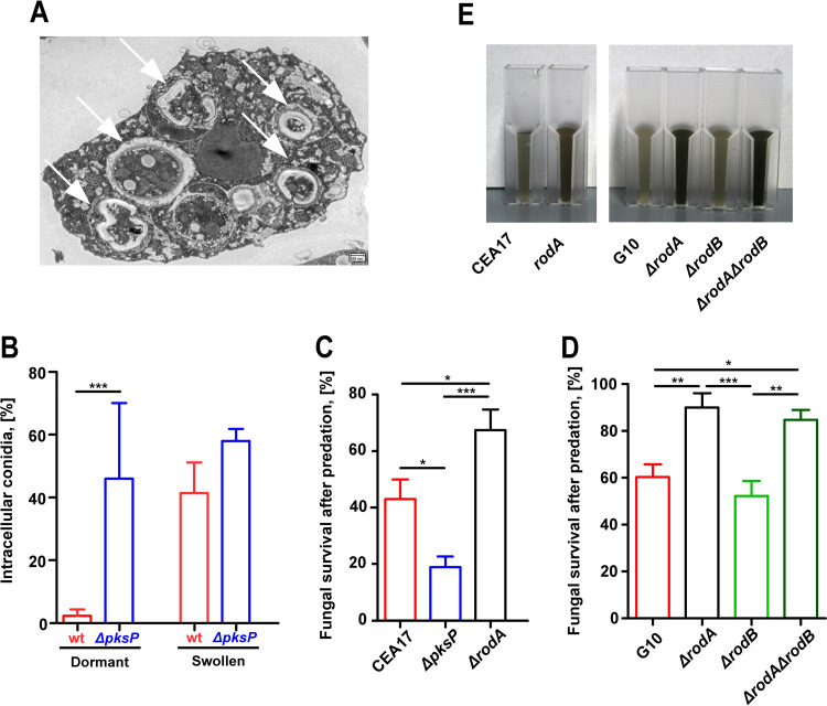
FIG 1.
Phagocytosis and killing of Aspergillus fumigatus by Protostelium aurantium. (A) Transmission electron micrograph (TEM) of P. aurantium with internalized conidia of A. fumigatus (white arrows) at different stages of digestion. (B) Uptake of dormant or swollen spores of A. fumigatus conidia after 1 h of coincubation by P. aurantium. wt, wild type. (C and D) Viability of conidia after P. aurantium predation. Fungal survival of the CEA17, ΔpksP, and ΔrodA strains (C) and the G10, ΔrodA, ΔrodB, and ΔrodA ΔrodB strains (D) was determined from resazurin-based measurements of fungal growth following confrontations with P. aurantium at an MOI of 10. Fungal survival data are expressed as means and standard errors of the means (SEM) of results from three independent experiments. Statistically significant differences were calculated with a Bonferroni posttest after a two-way ANOVA, with significance shown as follows: *, P < 0.05; **, P < 0.01; ***, P < 0.001. (E) Suspensions of 109 conidia of A. fumigatus strains showing different levels of DHN-melanin exposure. CEA17 and G10 represent wild type-like strains; ΔpksP is a deletion mutant for the first gene in 1,8-DHN-melanin biosynthesis; ΔrodA, ΔrodB, and ΔrodA ΔrodB are deletion mutants for genes encoding surface hydrophobins.

