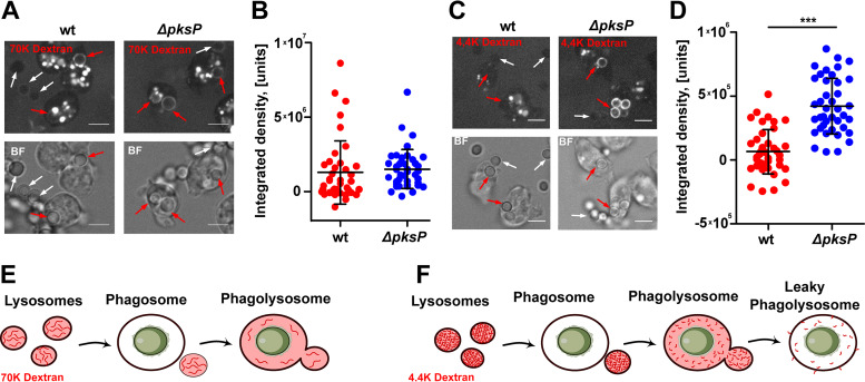
FIG 6.
Dextran leakage and Vps32 trafficking to conidium-containing phagolysosomes. (A and C) Cells of D. discoideum were loaded with fluorescent dextran at a molecular weight of 70,000 Da (A) or 4,400 Da (C) and subsequently infected with A. fumigatus conidia. Images were captured after 300 min p.i. Internalized conidia and free conidia are indicated by red and white arrows, respectively. (B and D) Quantification of red fluorescence of the two dextrans (70 kDa [B] and 4.4 kDa [D]) as integrated densities in conidium-containing phagosomes after 300 min p.i. Values were normalized by background subtraction of free conidia. Data are based on results from 3 biological replicates, with statistically significant differences calculated in a one-way ANOVA with P values of <0.0001. (E and F) Schematic representation of size-discriminated leakage of dextran from phagolysosomes for 70-kDa dextran (E) and 4.4-kDa dextran (F).

