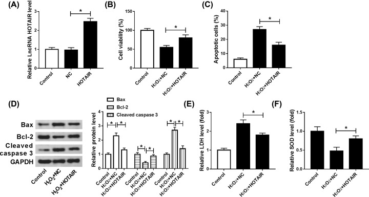
Figure 3. Up-regulation of HOTAIR induced cell viability, hindered apoptosis and decreased oxidative stress damage in H2O2-induced cardiomyocytes.
(A–F) The level of HOTAIR was determined by qRT-PCR (A) after transfected with HOTAIR or NC in H9c2 cells. (B–F) H2O2-induced cardiomyocytes were transfected with HOTAIR or NC, respectively. (B) Cell viability was assessed by CCK-8. (C) Flow cytometry was conducted to evaluate cell apoptosis in vitro. (D) Apoptosis-relative proteins of Bcl-2, Bax and Cleaved-caspase 3 were determined by Western blot. ELISA was carried out to examine the level of LDH and SOD; *P<0.05.

