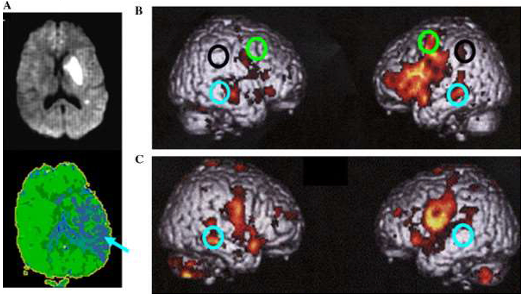
Fig. 3.

(A) DWI (top) done at Day 1 and PWI done at week 6 (bottom) of a patient with almost complete resolution of initial anomia and comprehension deficits, showing chronic hypoperfusion of left temporal cortex, including BA 37 (arrow). (B) fMRI study of word generation in six healthy control patients, showing significant BOLD effect in left BA 6 (dorsal posterior frontal; green circles), left BA 22, 37 (posterior temporal areas; blue circles), left BA 40 (supramarginal gyrus in the parietal lobe; black circles), and bilateral BA 44, 45 (posterior, inferior frontal) and 41/42 (auditory cortex), as well as right BA 21 (middle temporal gyrus). (C) fMRI study of word generation in the patient whose DWI and PWI are shown, demonstrating absence of the expected BOLD effect in the hypoperfused region (BA 37) and increased BOLD effect in the homologous region of the opposite hemisphere.
