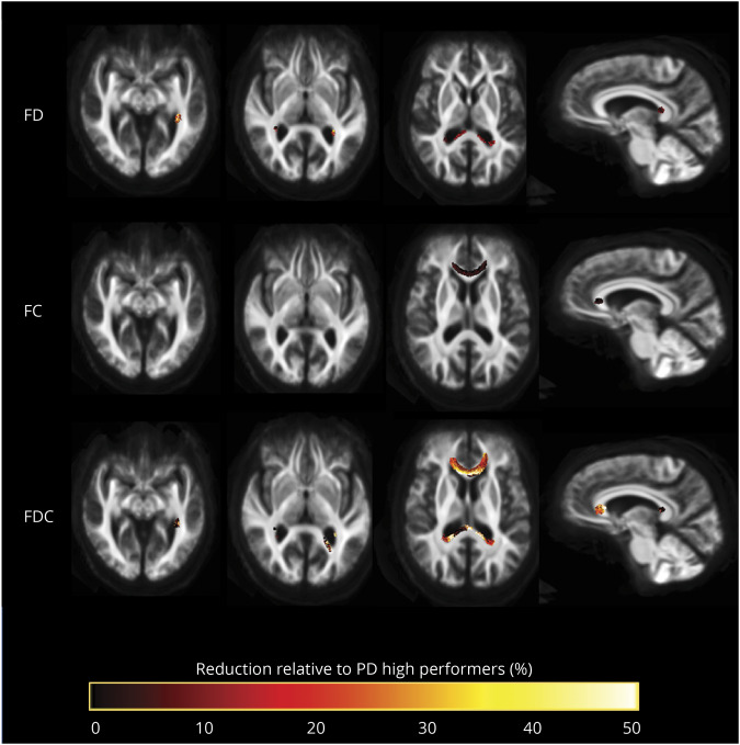
Figure 3. Fiber tract-specific reductions in PD low performers compared to PD high performers from whole-brain fixel-based analysis.
Patients with Parkinson disease (PD) and low visual performance showed macrostructural (changes in fiber cross section [FC]) and microstructural changes (changes in fiber density [FD]). Microstructural changes are seen within the splenium of the corpus callosum, posterior thalamic radiations bilaterally, and left inferior fronto-occipital fasciculus. Macrostructural changes are seen within the corpus callosum. Changes in the combined FD/FC (FDC) metric are seen in the genu, splenium, bilateral thalamic radiations, and left inferior fronto-occipital fasciculus; this represents impaired overall ability to relay information in these tracts in PD low performers. Results are displayed as streamlines; these correspond to fixels that significantly differed between PD low performers and PD high performers (family-wise error–corrected p < 0.05). Streamlines are colored by percentage reduction in the PD low performers group compared to high performers for FD, FC, and FDC.

