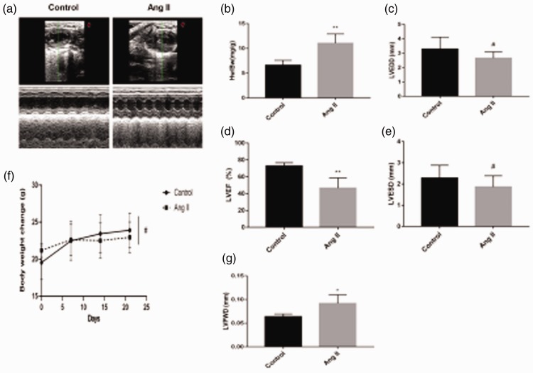
Figure 1.
(a) Cardiac structure detected by echocardiography. (b) Heart weight/body weight ratio of mice (**P < 0.01 versus the control group). (c) Left ventricular end-diastolic diameter (LVEDD) (#P > 0.05 versus the control group). (d) Left ventricular ejection fraction (LVEF) (%) of mice detected by echocardiography (**P < 0.01 versus the control group). (e) Left ventricular end-systolic diameter (LVESD) (#P > 0.05 versus the control group). (f) Changes in weight of mice were measured every week for 3 weeks (#P > 0.05). (g) Left ventricular posterior wall thickness (LVPWD) of mice detected by echocardiography (*P < 0.05 versus the control group). Ang II, angiotensin II.

