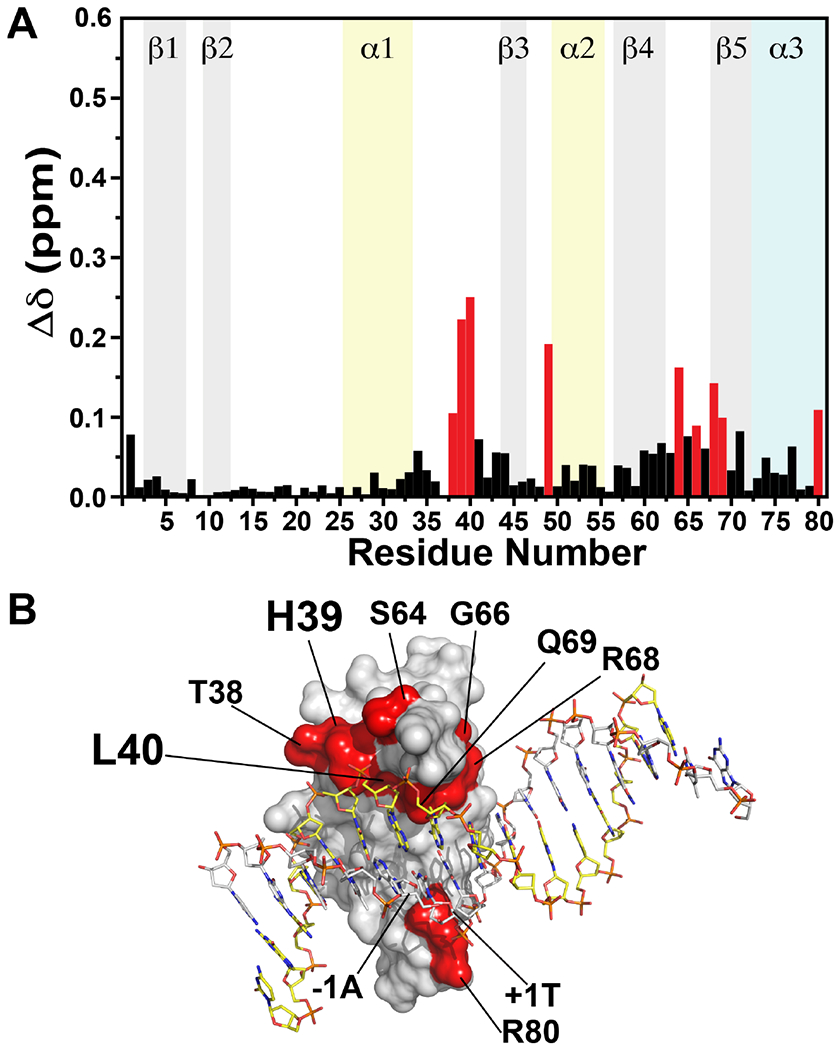
Figure 5.

(A) Differences in chemical shifts for TopN in the presence of two molar equivalents of spDNA or nsDNA. Resonances in the presence of spDNA were used as reference to calculate the perturbations using Equation 1. Residues with the largest differences are shown in red. (B) Residues with significant differences in resonance positions in the presence of spDNA or nsDNA are mapped onto the surface of the N domain in the crystal structure of of vTopIB bound to the consensus sequence, and colored red. H39 and L40 that exhibit the most significant differences are labeled in larger font. The C domain has been omitted for clarity.
