Figure 5.
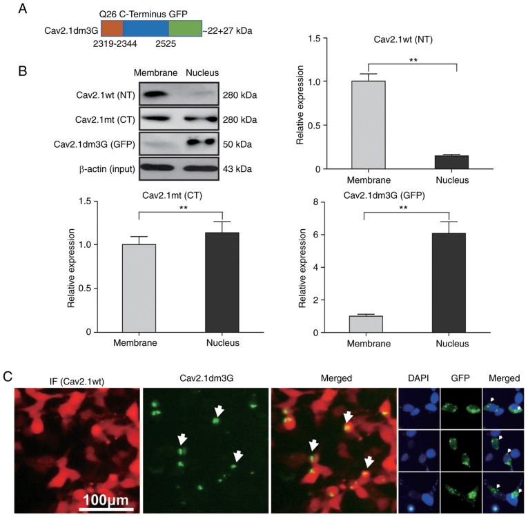
Cav2.1 mutant molecules are translocated to the nucleus of the SH-SY5Y cells. Plasmids expressing Cav2.1 wt, Cav2.1 mt and Cav2.1dm3G were transfected into SH-SY5Y cells, and proteins were collected from cell membranes or the nucleus and were detected by western blotting. (A) Diagram of the Cav2.1dm3-GFP fusion molecule. (B) Western blotting indicated that Cav2.1 wt molecules were located on the cell membrane, Cav2.1 mt molecules were located on both the cell membrane and in the cell nucleus, and Cav2.1dm3G molecules were translocated to the cell nucleus. β-actin was used to illustrate that the membrane proteins and nucleoproteins were extracted from the same quantity of transfected cells. Upper left panel, bands on the blotting membrane; the other panels, relative band density (t-test). (C) Immunofluorescence staining revealed the nuclear translocation phenomenon of Cav2.1 mt molecules. Cav2.1 mt, mutant-type Cav2.1; Cav.1 wt, wild-type Cav2.1; NT, N terminus; CT, C terminus. **P<0.01.

