Figure 1.
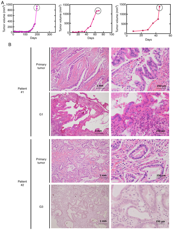
Establishment of pancreatic cancer PDX lines. (A) Tumor growth curves for a representative pancreatic PDX line. When the tumor volume reached ~1,000 mm3, the mouse was sacrificed, and the tumor was isolated and transplanted into another mouse. (B) Preserved morphological characteristics in xenograft tumors in NSG mice. H&E staining of the primary tumors and subsequent generations of PDXs for two patients. The pathological diagnosis of the primary tumors was tubular adenocarcinoma. PDX, patient-derived xenograft; H&E, hematoxylin and eosin.

