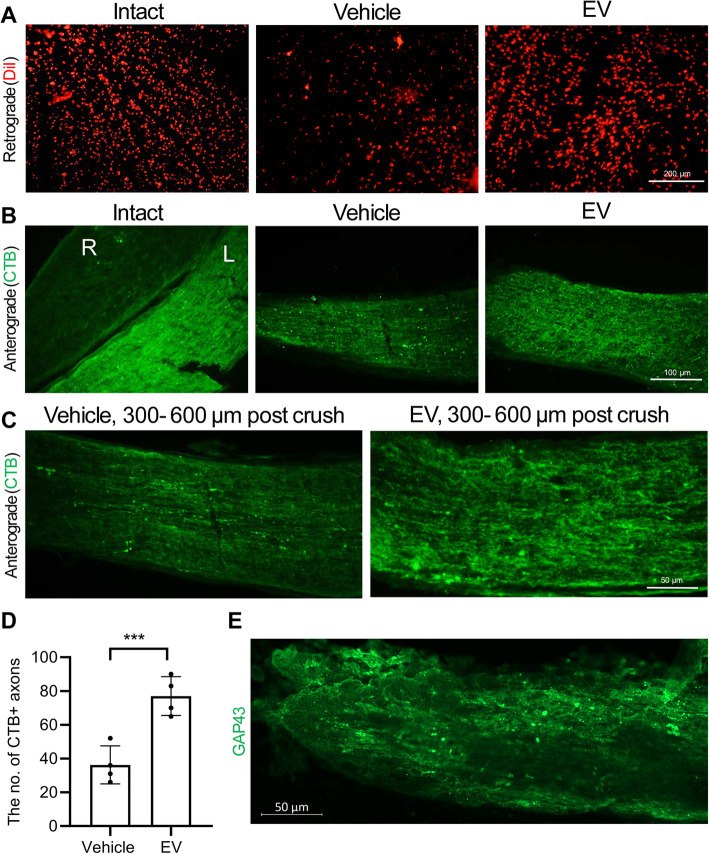
Fig. 4.
Retro- and anterograde axonal tracing 60 days post-treatment. a The intact retinal ganglion cells (RGCs) soma in the vehicle and extracellular vesicle (EV) groups that was retrogradely stained with DiIC18(3) (DiI; red). b, c Longitudinal cryosections and immunostaining against chlorotoxin B (CTB) in the intact group of the anterograde shows the left (L) and right (R) optic nerves. CTB was only injected in the left optic nerve. Data from the anterograde tracing shows that more CTB+ axons extended the length of the optic nerve in the EV group, whereas smaller numbers in the vehicle were seen, even at a distance of 300 to 600 μm in distal site of crush. d Quantification of CTB+ RGCs axons in the optic nerve in (c). Data are shown as mean ± SD for four optic nerves per group. The data were analyzed by the un-paired t test. ***P < 0.001. e A longitudinal section of the optic nerve with EV, which shows numerous axons with GAP43 expression

