Figure 2.
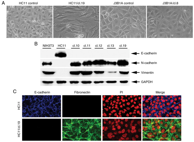
VL30 retrotransposition induces a mesenchymal phenotype associated with the protein expression of EMT markers in HC11 cells. (A) Fields of control HC11 or J3B1A cells and respective retrotransposition-positive clone cells (magnification, ×20) grown in normal culture dishes. HC11/cl.19 and J3B1A/cl.8 panels represent cells with an induced and non-induced mesenchymal phenotype, respectively. (B) Western blot analysis of whole protein lysates from NIH3T3, HC11 and HC11 retrotransposition-positive clone cells. Arrows indicate E-cadherin-, N-cadherin- and Vimentin-antibody reactions. GAPDH refers to sample protein load. (C) Immunofluorescence of control HC11 and HC11/cl.19 cells after staining with E-cadherin and fibronectin antibodies as well as propidium iodide (PI). Scale bar, 20 µm. Data in (B and C) are representative of 3 experiments.

