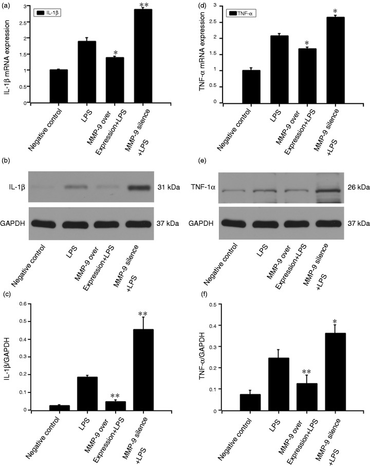
Figure 3.
Effect of MMP9 on LPS-induced expression of IL-1β and TNF-α. MC3T3-E1 cells were pre-treated with MMP9 overexpression plasmid or si-MMP9-3 (target sequences: GGAACTCACACGACATCTT) for 24 h. The 20 μg/ml LPS was added to the culture medium for another 12 h. qRT-PCR and Western blot were performed to detect IL-1β (a–c) and TNF-α (d–f) expressions. Quantification of protein expression was normalized to GAPDH using a densitometer (imaging system). The data are representative of three independent experiments and expressed as the mean ± SD. *P < 0.05 vs. LPS; **P < 0.01 vs. LPS.

