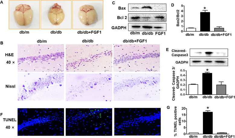
Fig. 4.
FGF1 blocks diabetes-induced morphological structure change and neuronal apoptosis in the hippocampus during DICD. a Morphological appearance of brains from db/m mice, db/db mice and db + FGF1-treated mice; b The H&E staining and Nissl staining of CA1 in the hippocampus from mice (Scale bar = 15 μm); c-e Western blotting and quantitative analysis of Bax, Bcl2 and Cleaved-Caspase3 expressions in the hippocampus from mice; f Representative images of TUNEL staining showing apoptotic cells (green signal) in the CA1 of hippocampus, cell nuclei were stained with DAPI (blue) (Scale bar = 15 μm); (g) The quantification of TUNEL-positive cells in the CA1 of hippocampus from mice. *p < 0.05, ***p < 0.001 vs. the other two groups, n = 3

