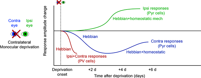
Figure 6. Time Course of Synaptic Changes Following Visual Deprivation.
Timeline of changes during short periods around MD. Initial firing rates of excitatory L2/3 neurons fall for the deprived contralateral eye on a short timescale, but partially recover over 1 week (Frenkel and Bear, 2004; Mrsic-Flogel et al., 2007). The initial loss of responsiveness is attributed to Hebbian mechanisms, but the slower recovery involves some homeostatic changes as well. In contrast, non-deprived eye responses strengthen more slowly. The transient rapid change is associated with the loss of excitation to PV+ interneurons, which occurs as rapidly as 1 day and is associated with the loss of excitatory synaptic input to these cells (Kuhlman et al., 2013).

