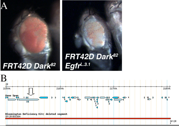
Figure 1.
A. Mosaic (FRT42D Dark82), and Dark82 EgfrL.3.1 (FRT42D Dark82EgfrL.3.1) eyes. In both eyes the homozygous mutant tissue is pigmented (w+mC). B. Region of chromosome 2R that failed to complement L.3.1 by deficiency mapping (2R:21,497,290..21,806,350). Arrow denotes location of Egfr gene. B is adapted from flybase.org (Gramates et al., 2017).

