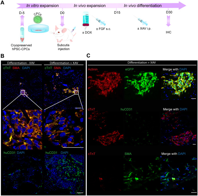
Figure 2.
CPC differentiation in vivo promoted by WNT-inhibition after subcutis injection. (A) Schematic of the experimental workflow for CPC expansion after subcutaneous injection in mouse, followed by directed differentiation by intraperitoneal (i.p.) injection of XAV939. (B) Human cTnT, SMA and human-specific CD31 (huCD31) immunostaining in plugs from mouse subcutis after the CPC expansion and differentiation protocol with or without i.p. injection of XAV939; scale bar = 100 µm in upper panel and lower panel; scale bar = 25 µm in middle panel. (C) Additional immunostaining with human-specific antibodies after CPC differentiation in the subcutis in the presence of XAV939: alpha-actinin is shown to co-label with eGFP in CMs; cTnT+ areas are shown with interspersed huCD31+ endothelial cells (ECs); and SMA+ cTnT-labelling indicates the presence of SMCs; scale bar = 25 µm.

