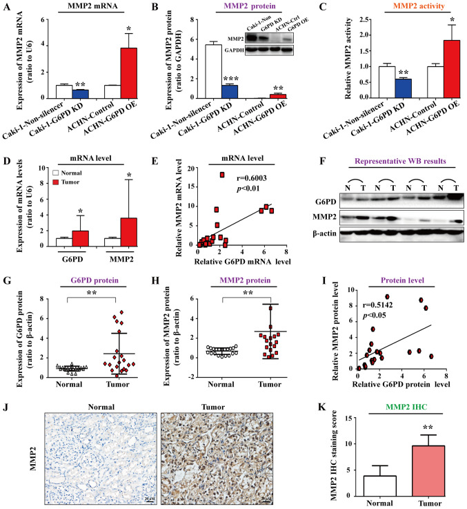
Figure 2.
G6PD promoted and was positively correlated with MMP2 expression in ccRCC cells. MMP2 mRNA and protein expression in stable transfected Caki-1 or ACHN cells were analyzed by (A) RT-qPCR and (B) western blotting, respectively. (C) MMP2 activities in stable transfected Caki-1 or ACHN cells was detected by assay kit, and raw data (fluoresence intensity) were transferred into the relative activity changes compared with each control. Evaluation of G6PD and MMP2 expression was conducted in 20 pairs of ccRCC tumor specimens and matched adjacent normal tissues by using (D) RT-qPCR and (F-H) western blotting. Pearson correlation analysis between (E) MMP2 and G6PD mRNA expression and (I) MMP2 and G6PD protein expression. Each analysis was performed at least three times and each sample assessed in triplicate. Data were expressed as the means ± standard deviation. *P<0.05, **P<0.01 and ***P<0.001 vs. non-silencer, control or adjacent normal tissues. (J) IHC analysis of MMP2 expression in 20 pairs of ccRCC specimens and matched adjacent normal tissues. Scale bar, 20 µm. (K) Quantification of the MMP2 staining score from (J). **P<0.01 vs. normal tissues. ccRCC, clear cell renal cell carcinoma; G6PD, glucose-6-phosphate dehydrogenase; IHC, immunohistochemistry; KD, knockdown; OE, overexpressing; RT-qPCR, reverse transcription quantitative PCR

