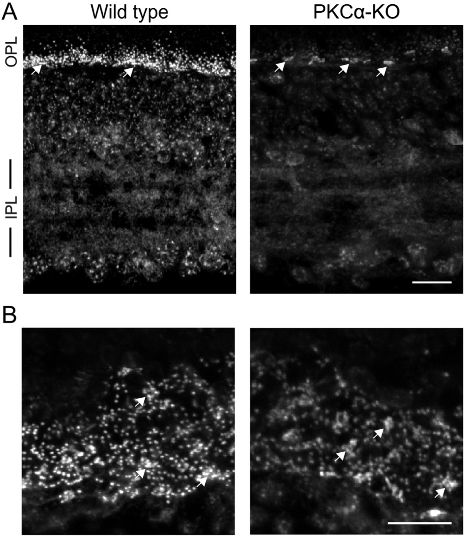
Fig. 1.

Phosphoserine labeling in the OPL is reduced in PKCα-KO retina. (A) Immunofluorescence confocal images of mouse retina sections from wild type and PKCα-KO retinas labeled with an antibody against phosphoserine residues within canonical PKC motif phosphoserine (PKC motif p-serine). (B) Images of PKC motif p-serine immunofluorescence in the outer plexiform layer of wild type and PKCα knockout (KO) retinas in obliquely cut sections. In both A and B, white arrows indicate labeling associated with presumptive cone pedicles. Scale bars: 20 μm. OPL: outer plexiform layer; IPL: inner plexiform layer.
