Figure 2.
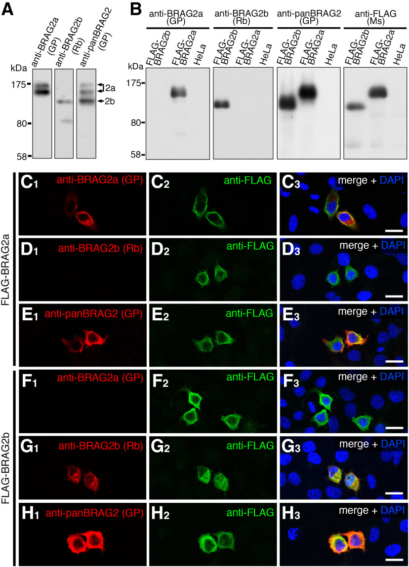
Characterization of BRAG2 antibodies. A, Immunoblotting of mouse brain lysates using guinea pig (GP) anti-BRAG2a, rabbit (Rb) anti-BRAG2b, and GP anti-panBRAG2 antibodies. Note that anti-panBRAG2, anti-BRAG2a, and anti-BRAG2b antibodies recognize three bands (160, 140, and 110 kDa), two bands (160 and 140 kDa), and a single band (110 kDa), respectively. B, Immunoblotting of HeLa cells transfected with FLAG-BRAG2a or FLAG-BRAG2b using anti-BRAG2a, anti-BRAG2b, anti-panBRAG2, and mouse (Ms) anti-FLAG antibodies. Note the specificity of anti-BRAG2a and anti-BRAG2b antibodies without cross-reactivity with FLAG-BRAG2b and FLAG-BRAG2a, respectively. C–H, Double immunofluorescence of HeLa cells transfected with FLAG-BRAG2a (C–E) or FLAG-BRAG2b (F–H) using anti-FLAG (C2–H2) and anti-BRAG2a (C1, F1), anti-BRAG2b (D1, G1), or anti-panBRAG2 (EI, H1) antibodies. Note the absence of the cross-reactivity of anti-BRAG2a and anti-BRAG2b antibodies with FLAG-BRAG2b and FLAG-BRAG2a, respectively. HeLa cells were counterstained with DAPI. Scale bars, 10 μm.

