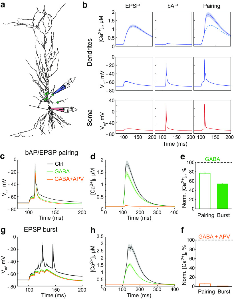
Figure 3.
Tonic GABAA conductances differentially affect responses to bAP/EPSP pairing and EPSP burst in the model pyramidal neuron. a, The synapse location (green dots) on the simulated pyramidal neuron. Two electrodes indicate where Vm was obtained: red, on soma; blue, on a second-order dendritic branch. The somatic electrode also indicates where the current injection used to elicit bAP occurred. b, The Ca2+ transients in the dendrite (top row); Vm in the dendrite (middle row) and the soma (bottom row). Left column, EPSP. Middle column, bAP. Right column, bAP/EPSP pairing. [Ca2+]i, Intracellular Ca2+ concentration. c, The ΔVm to bAP/EPSP pairing in control (Ctrl, gray), on an increase in the tonic GABAA conductance (GABA, green), and subsequent blockade of NMDARs (GABA + APV, orange). d, The Ca2+ transient in response to bAP/EPSP pairing in control (gray), and on an increase in the tonic GABAA conductance (green), and subsequent blockade of NMDARs (orange). e, The summary of data on the effect of tonic GABAA conductances (GABA) on the amplitude of the Ca2+ transient induced by bAP/EPSP pairing (empty bar) and EPSP burst (filled bar). f, The summary of data on the effect of subsequent NMDAR blockade (GABA + APV) on the amplitude of the Ca2+ transient induced by bAP/EPSP pairing (empty bar) and EPSP burst (filled bar). g, The ΔVm to EPSP burst in control (gray), and on an increase in the tonic GABAA conductance (green), and subsequent blockade of NMDARs (orange). h, The Ca2+ transient in response to EPSP burst in control (gray), and on an increase in the tonic GABAA conductance (green), and subsequent blockade of NMDARs (orange).

