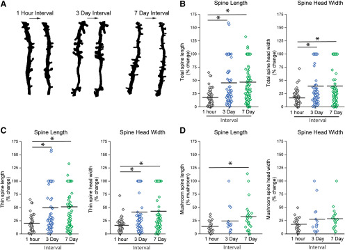
Figure 3.
Normal dendritic spine length and head width dynamics within the dorsal horn of the spinal cord. Absolute % change in (B–D) spine length and head width (between time intervals of 1 h, 3 d, and 7 d). A, Representative images of in vivo paired images with intervals of 1 h, 3 d, and 7 d. B, Absolute % change in length and head width of all spine types (total spines). Dynamics within the total spine population over a 1 h period were less compared with the dynamics observed over 3 and 7 d intervals. C, Absolute % change in length and head width of thin spines. There was a greater change in length and head width of thin spines over 3 and 7 d time intervals, which was not observed during a single hour. D, Absolute % change in length and head width of mushroom spines. Mushroom-shaped spines are stable over a 3 d interval. Over a 7 d interval, there was an increase in the absolute % change in length of mushroom spines. *p < 0.05 (one-way ANOVA, Kruskal–Wallis test).

