Figure 4.
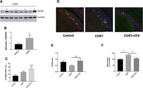
CORT increases BCAN and PNN levels. A, B, BCAN levels in hippocampal lysates from control and CORT-exposed mice (Western blot and densitometry, respectively). BCAN levels are increased in hippocampal lysates from CORT-exposed mice compared with controls (n = 4 mice per group, p = 0.0466, Student's t test). C, Ratios of MMP-9 and its inhibitor, TIMP-1, in hippocampal lysates from CORT-exposed animals treated with saline or VFX. VFX significantly increased MMP-9/TIMP-1 levels over control (n = 5 or 6 mice per group, p = 0.0027, one-way ANOVA with Tukey's multiple comparisons), whereas CORT alone had no significant effect compared with control (p = 0.094, one-way ANOVA with Tukey's multiple comparisons). D, Representative PNN staining for control, CRT, and CORT + VFX exposed WT animals. E, F, Quantification of the PV/PNN ratio and PNN intensity. VFX significantly increases the PNN/PV ratio in CORT-exposed animals (6-8 mice, n = 18-26 slides, p = 0.0383, ANOVA with Tukey's multiple comparisons) and reduces overall PNN fluorescence intensity (p < 0.0008), consistent with a reduction in PNN levels. Scale bar, 250 μm. *p < 0.05, **p < 0.005.

