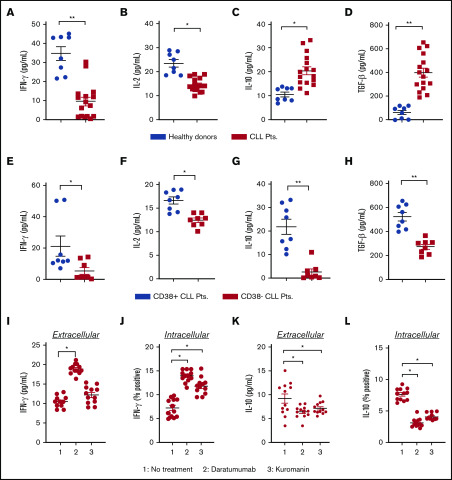
Figure 1.
Effect of anti-CD38 agents on the level of pro and anti-inflammatory cytokines in patients. (A-D) Plasma from healthy donors (n = 8) or patients with CLL (n = 16) were isolated from blood, and concentration of IFN-γ (A) and IL-2 (B) was measured by sandwich enzyme-linked immunosorbent assay. Similarly, levels of IL-10 (C) and TGF-β (D) were assessed. From the overall data set, subset analysis of IFN-γ (E), IL-2 (F), IL-10 (G), and TGF-β (H) levels in patients with clinically defined CD38+ (n = 8) and CD38− (n = 8) disease was conducted. (I-J) The effect of daratumumab (1 µg/mL, 72 hours) or kuromanin (30 µM, 72 hours) on extracellular (I) IFN-γ from patients with CLL was examined (n = 12, n = 3 with CD38− disease and n = 9 with CD38+ disease). After drug treatment with daratumumab or kuromanin, supernatant from the PBMCs of patients with CLL was collected via centrifugation and cytokines measured by sandwich enzyme-linked immunosorbent assay. (J) Intracellular IFN-γ positivity in CD19+ gated CLL cells (from the same patients as in panel I) was measured after treatment with daratumumab or kuromanin (same concentrations/time as in panel I); 4 hours before termination of experiments, ion channels were blocked with ionomycin and brefeldin used for GolgiStop. Extracellular IL-10 (K) and IL-10+ (L) CLL cells were measured in the same manner as in panels I and J, respectively. For extracellular cytokines, data were derived from a standard curve and represented as picograms per milliliter ± SEM in a scatter dot plot for each set of experiments. For intracellular staining, data were represented as percentage positive cells ± SEM. Each experiment was performed at least thrice in duplicate. *P < .05; **P < .001.

