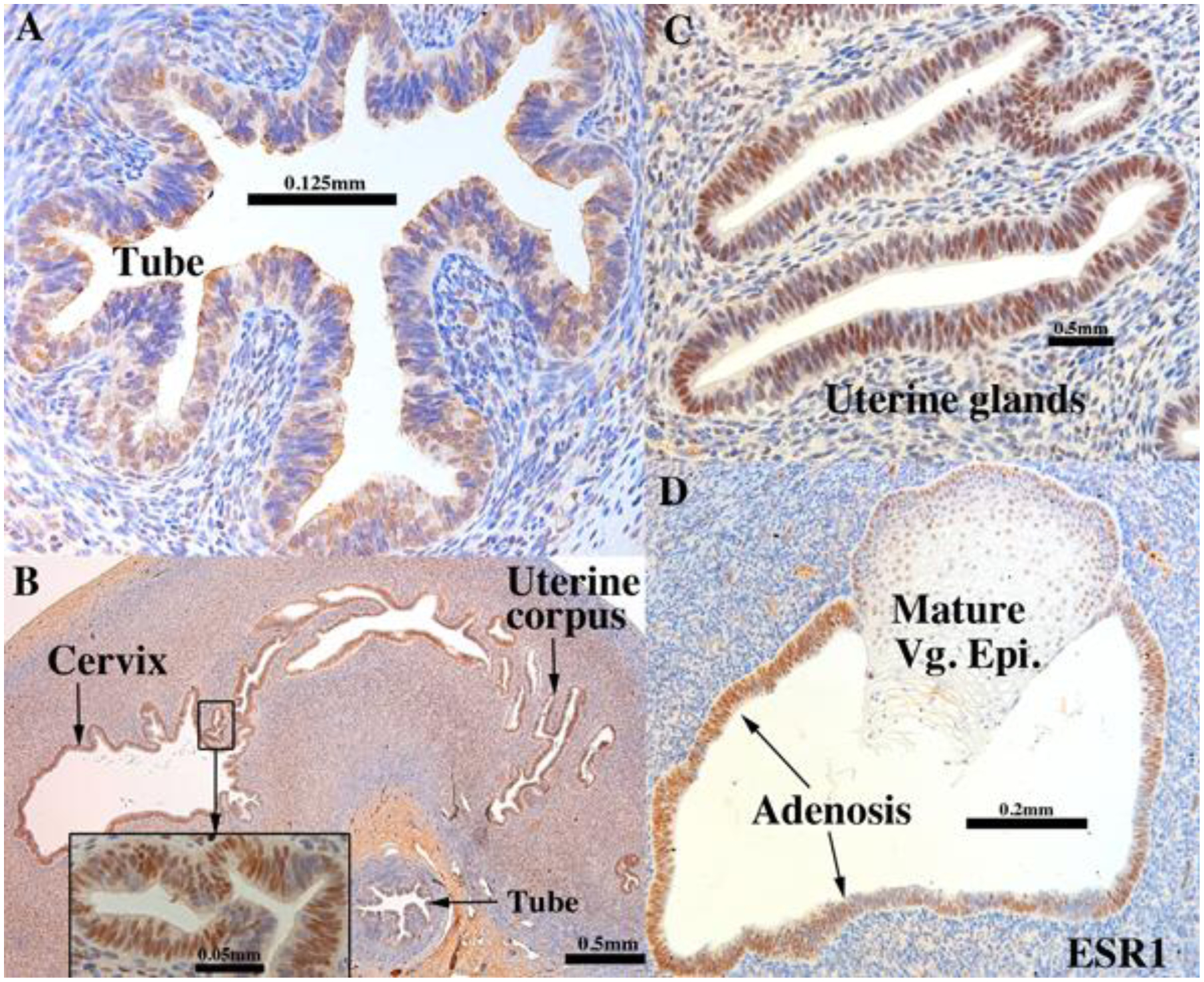
Figure 10.

ESR1 immunostaining in an intact 13-week human fetal reproductive tract (AC122) grown for 4 weeks in a DES-treated ovariectomized female athymic mouse host. Tubal epithelium (A) expresses ESR1 but at a level reduced relative to non-grafted and specimens grafted into untreated hosts. ESR1 expression in the uterine tube, glands of the uterine corpus and cervix (B) (low magnification}, (C) (high magnification) of uterine glands. In the vaginal region ESR1 reactivity is seen in both mature squamous epithelium and adenosis, while the mesenchyme is largely unreactive (D).
