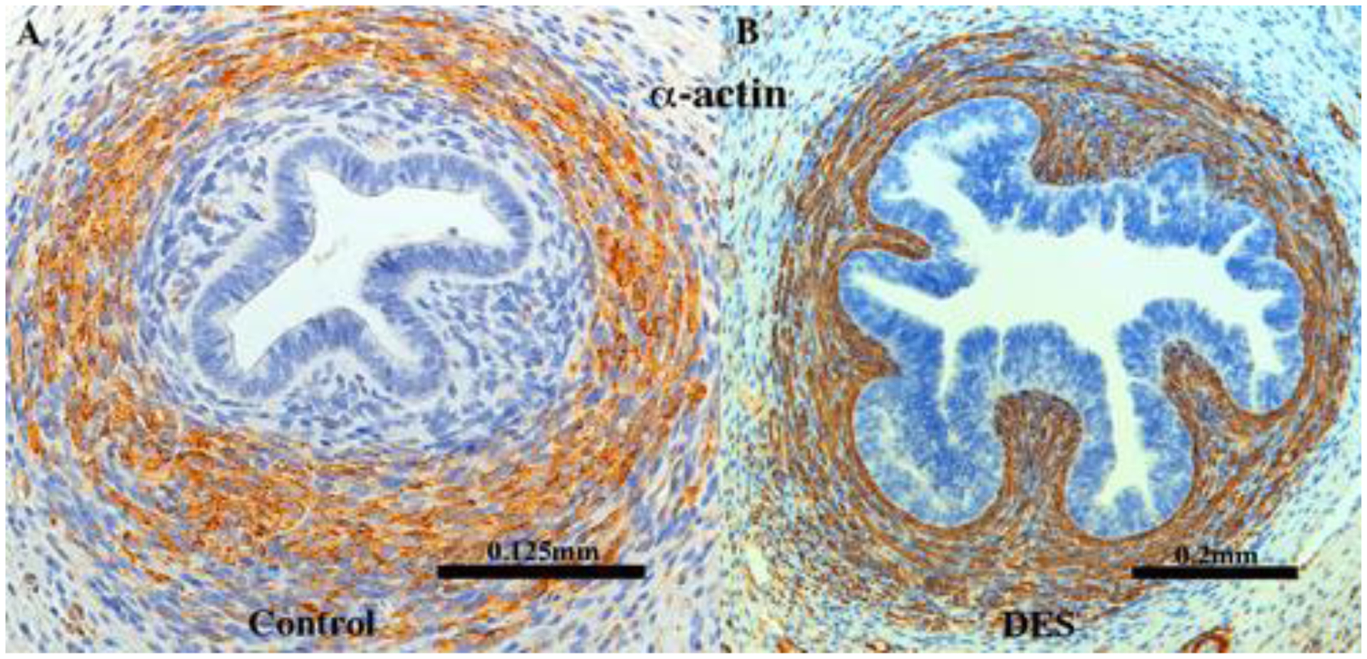
Figure 19.

αACT2 immunostaining of 13-week human uterine tubes grown for 4 weeks in (A, [AC121]) an untreated ovariectomized host and (B, [AC122]) in a DES-treated ovariectomized host stained for αACT2. Note that in the control (A), a substantial mesenchymal layer encompasses the tubal epithelium, which DES treatment (B) has obliterated, resulting in a misplaced αACT2 smooth muscle layer, which now comes into a intimate association with the epithelium.
