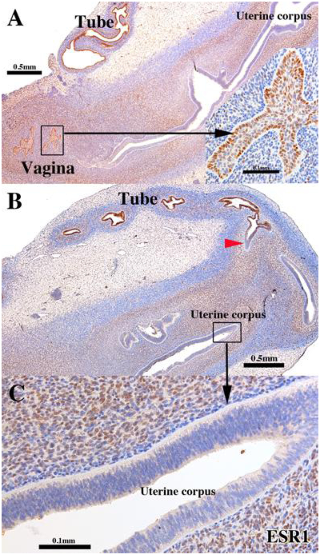
Figure 2.

ESR1 immunostaining of a 13-week intact female fetal reproductive tract (AC121) grown for 4 weeks in an untreated ovariectomized female athymic mouse host. The epithelia of the uterine tube and vaginal plate (UGS-derived) are reactive for ESR1, while endometrial/cervical epithelia are not (B). The red arrowhead marks the uterotubal junction remarkable for the sharp transition in ESR1 staining. Detail of ESR1-negative endometrial epithelium (C). Note that the inner layer of mesenchymal cells of the uterine corpus (presumed endometrial stroma) is strongly ESR1-positive in contrast to peripheral mesenchyme (presumptive myometrium), which is weakly ESR1-positive. The stroma of the uterine tube is also ESR1 reactive but to a far lesser degree than in the corresponding area of uterine corpus.
