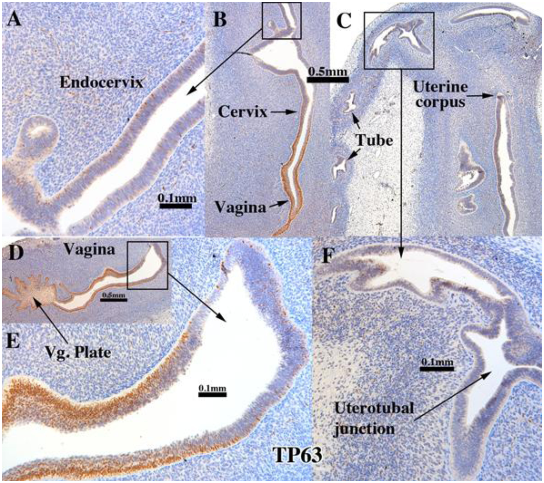
Figure 7.

TP63 immunostaining of an intact 13-week human fetal reproductive tract (AC121) grown for 4 weeks in an untreated ovariectomized female athymic mouse host. TP63 was strongly expressed in the stratified epithelium of the vaginal plate (D), vagina (B & D), but not in the simple columnar epithelia of the uterine tube (C), uterotubal junction (F) and upper uterine. Transitions between stratified and simple columnar epithelium (D-E) exhibited a sharp reduction in TP63 to patchy single cell expression, which was also seen in the presumed endocervical region (A).
