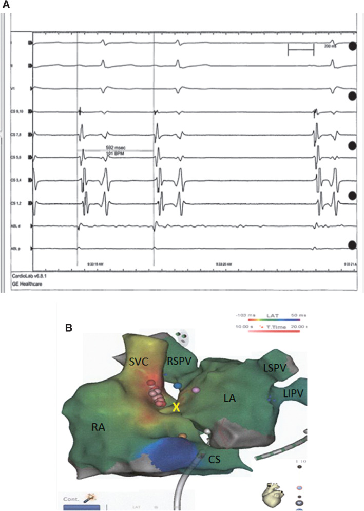
Figure 2:
A: Intracardiac electrogram revealing slowing and termination of the tachycardia with ablation of the RSPV. B: CARTO® 3 (Biosense Webster, Diamond Bar, CA, USA) map of the RA and LA in an anteroposterior top-down position. In the LA, the blue tag represents an ablation point adjacent to the RSPV with termination. In the RA, multiple ablation tags (pink and red) represent ablation points in the posteroseptal area and termination with the red tag. The yellow tag represents the His bundle, whereas the yellow X denotes the transseptal area. RA: right atrium; SVC: superior vena cava; RSPV: right superior pulmonary vein; LSPV: left superior pulmonary vein; LA: left atrium; LIPV: left inferior pulmonary vein; CS: coronary sinus.

