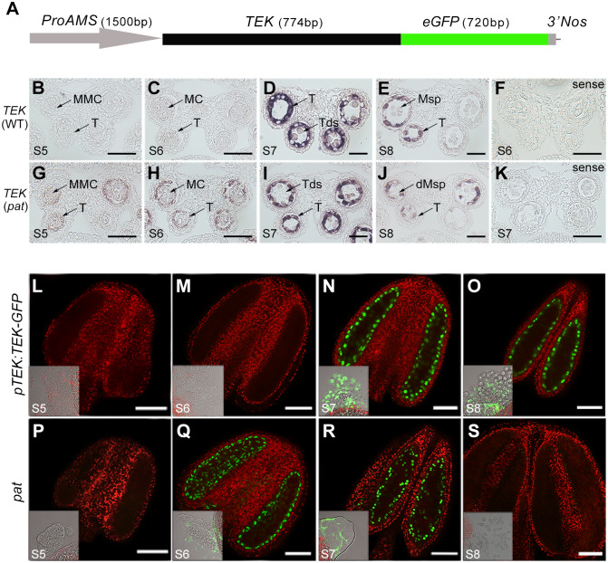
Fig 1. Precocious expression of TEK transcripts and TEK-GFP proteins driven by the AMS promoter in pat.
(A) The pAMS:TEK–GFP constructs included a 1500 bp AMS promoter, TEK genomic fragment and GFP coding region. RNA in situ hybridization of TEK transcript in WT (B–E) and pat (G–J) anthers at stages 5–8 was performed using an antisense probe. TEK transcript in WT anthers at stage 6 (F) and pat anthers at stage 7 (K) using a sense probe. MMC, microspore mother cell; MC, meiocytes; T, tapetum; Tds, tetrads; Msp, microspore. Scale bars, 20 μm. Fluorescence confocal images of the TEK–GFP fusion protein in the anthers of pTEK:TEK-GFP transgenic plants (L-O) and pat (P-S) at stages 5–8. TEK-GFP was specifically located in the tapetal nuclei and expressed at stages 7–8 in pTEK:TEK-GFP transgenic anthers (N and O), while in pat anthers this protein was precociously expressed at stages 6–7 (Q and R). The bright-field images are located at the bottom left, showing that GFP fluorescence was observed only in the tapetal cells. Scale bars, 50 μm.

