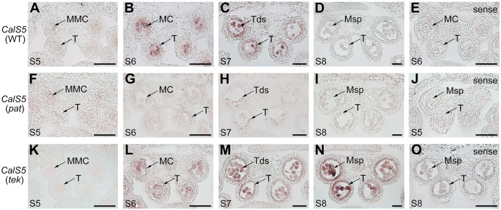
Fig 5. CalS5 expression patterns in the wild-type, pat and tek anthers.
Expression of CalS5 in microspore mother cells, tetrads and tapetum was tested by RNA in situ hybridization in WT (A–D), pat (F-I) and tek anthers (K-N) at stages 5–8 using an antisense probe. According to the stages at which CalS5 reaches its peak with an antisense probe, its transcript was observed with a sense probe in WT anthers at stage 6 (E), pat anthers at stage 5 (J), and tek anthers at stage 8 (O). MMC, microspore mother cell; MC, meiocytes; T, tapetum; Tds, tetrads; Msp, microspore. Scale bars, 20 μm.

