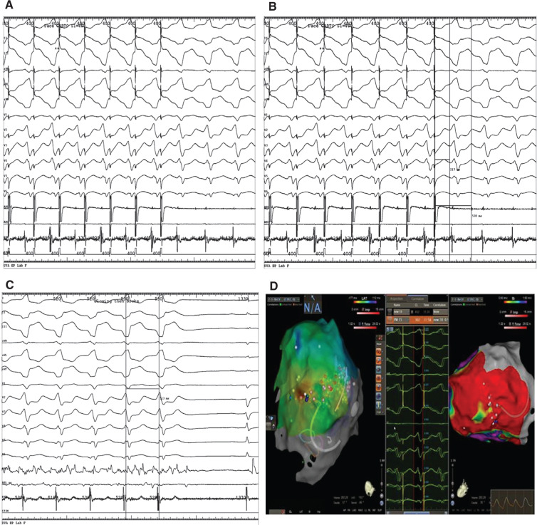
Figure 3:
A: Entrainment of ventricular tachycardia, showing concealed fusion. B: Entrainment with a PPI TCL of ~30 ms to 40 ms with stim-QRS~EGM-QRS. Stim-QRS in 30% to 70% of the TCL. C: Slowing and then termination of the tachycardia after RF ablation. D: Electroanatomic maps showing early activation areas and areas of scar on bipolar mapping on the right.

