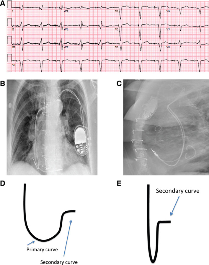
Figure 2:
A: A 12-lead ECG with atrial and RV low septal pacing; QRS duration (QRSd): 150 ms. Note the Q wave in leads I and aVL. B: A posterioanterior view of the right atrial and septal RV leads on chest X-ray. C: A lateral view of the right atrial and septal RV leads. D: Schematic representation of the stylet shape showing the primary and secondary curves. E: Foreshortened view of the stylet shape demonstrating the secondary curve.

