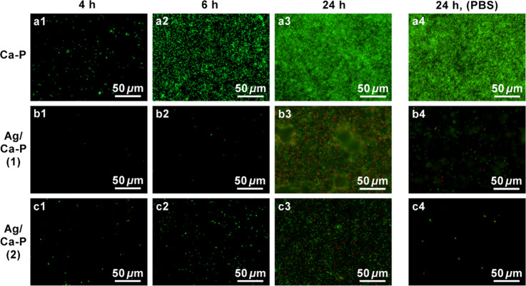
Figure 7.
Fluorescence microscopy images of S. aureus on (a) Ca-P coating as control, (b) Ag/Ca-P(1) coating, and (c) Ag/Ca-P(2) coating after incubation for (a1–c1) 4 h, (a2–c2) 6 h and (a3–c3) 24 h, and (a4–c4) after 24 h incubation on the coatings pretreated by PBS solution. Green and red indicate live and dead bacteria, respectively.

