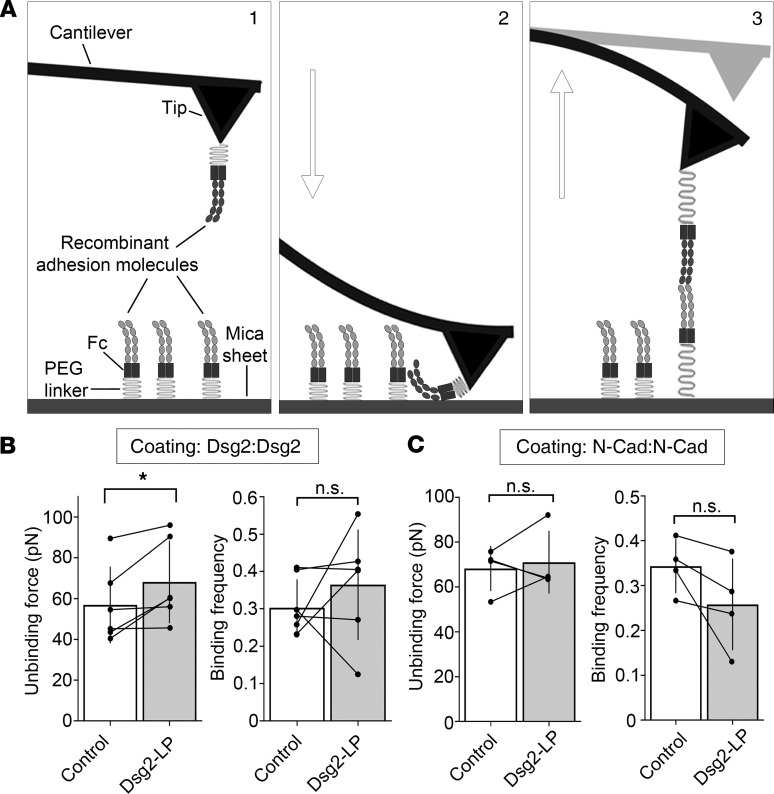
Figure 5. Dsg2-LP stabilizes Dsg2 binding in vitro.
(A) Schematic of atomic force microscope (AFM) force mapping setup. The tip of a cantilever functionalized with the extracellular domains of adhesion molecules via a flexible PEG linker (step 1) is repeatedly extended to (step 2) and retracted from (step 3) a functionalized surface. Interaction of the tip- and surface-bound proteins is detectable by cantilever deflection. (B and C) Analysis of unbinding force and binding frequency of cell-free AFM measurements with Dsg2- or N-Cad–coated tips probed on mica sheets with respective coatings. Every dot in the right panel represents the mean value of 1 independent experiment, bars indicate mean ± SD, and black lines connect paired values. n = 6 for Dsg2; n = 5 for N-Cad. n.s., P ≥ 0.05, *P < 0.05. Two-tailed paired Student’s t test.

