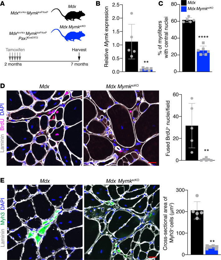
Figure 1. Genetic deletion of Myomaker in SCs of mdx mice leads to complete loss of fusion.
(A) Schematic showing the mouse model and timing of tamoxifen injections. (B) qPCR analysis of Myomaker from whole diaphragm indicates that Myomaker is significantly reduced in mdx MymkscKO muscle. (C) Percentage of myofibers with central nucleation in laminin and DAPI-stained tibialis anterior (TA) sections. (D) Mice were given intraperitoneal injections of BrdU for 7 days before sacrifice to label proliferating nuclei. BrdU+DAPI+ nuclei within the borders of laminin-labeled myofibers were defined as fused nuclei. Quantification shows loss of fusion in mdx MymkscKO mice. (E) Immunofluorescence staining for Myh3+ (embryonic myosin) myofibers reveals loss of de novo myofiber formation in mdx MymkscKO mice. Quantification of cross-sectional area of Myh3+ cells supports lack of true myofiber formation. Statistical analyses and data presentation: (B, D, and E) Mann-Whitney U test; **P < 0.01; (C) unpaired t test; ****P < 0.0001. Data are represented as mean ± SD. Scale bar: 20 μm. n = 5–6.

