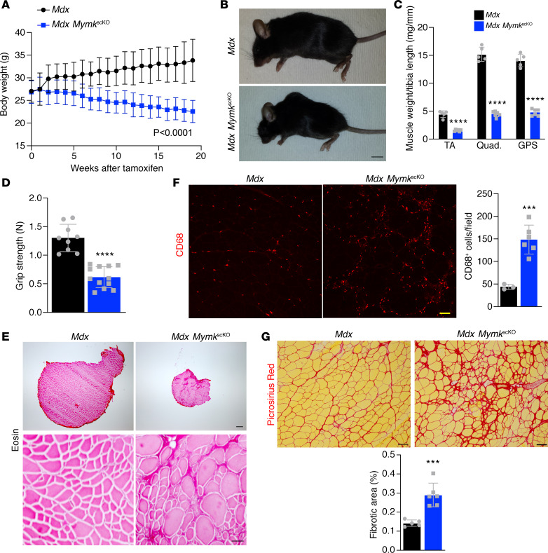
Figure 2. Deletion of Myomaker in SCs of dystrophic muscle leads to severe pathology.
(A) Body weight was assessed weekly and dropped steadily following loss of Myomaker in SCs (n = 9–12). (B) Mdx MymkscKO show loss of muscle mass and kyphotic spinal morphology by gross examination. (C) Dry weights of tibialis anterior (TA), quadriceps, and gastrocnemius/plantaris/soleus (GPS) following dissection. (D) Muscle function was assessed using a forelimb grip strength meter (n = 9–12). (E) Eosin stains on cross section of TA muscles show reduced muscle cross-sectional area and altered myofiber morphology. (F) CD68 immunofluorescence of TA sections shows enhanced inflammatory process in mdx MymkscKO muscle (n = 3–6). (G) Picrosirius red–stained sections of TA muscle demonstrate increased fibrosis in fusion-incompetent dystrophic muscle. Statistical analyses and data presentation: (A) linear regression with slopes comparison; (D, F, and G) unpaired t test; ***P < 0.001, and ****P < 0.0001. Data are represented as mean ± SD. Scale bars: 1 cm (B), 200 μm (top right) and 50 μm (bottom right) (E), 50 μm (F), 100 μm (G). n = 5–6 except where noted.

