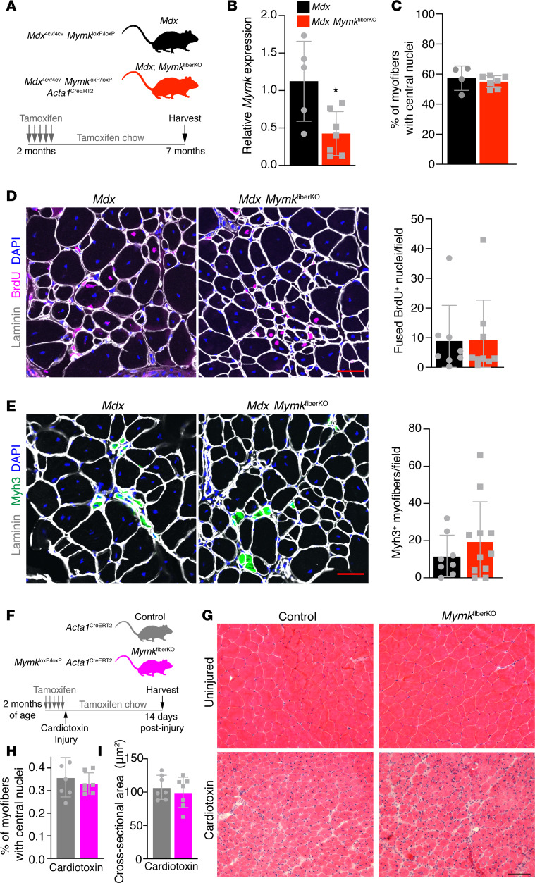
Figure 4. Deletion of Myomaker in mature myofibers does not affect fusion dynamics in WT or mdx mice.
(A) Schematic showing the mouse model and the timing of tamoxifen administration. (B) qPCR analysis from whole diaphragm muscle indicates reduced Myomaker expression in mdx MymkfiberKO mice (n = 5–7). (C) Quantification of centrally nucleated myofibers in tibialis anterior (TA) muscle revealed no change following deletion of Myomaker in myofibers (n = 4–7). (D) Fusion of BrdU+ nuclei is unaffected in mdx MymkfiberKO mice (n = 8–11). (E) Formation of de novo (Myh3+) myofibers is not significantly altered in mdx MymkfiberKO muscle (n = 8–11). (F) Schematic showing acute injury mouse model, timing of cardiotoxin injury, and tamoxifen regimen. (G) H&E stain of TA muscle 14 days postinjury. (H) Quantification of myofibers with central nuclei from laminin-stained sections (not shown) (n = 7). (I) Average cross-sectional area of TA myofibers was unchanged following Myomaker deletion in myofibers (n = 7). Statistical analysis and data presentation: (B) unpaired t test, *P < 0.05. Data are represented as mean ± SD. Scale bars: 50 μm (D and E), 100 μm (G).

