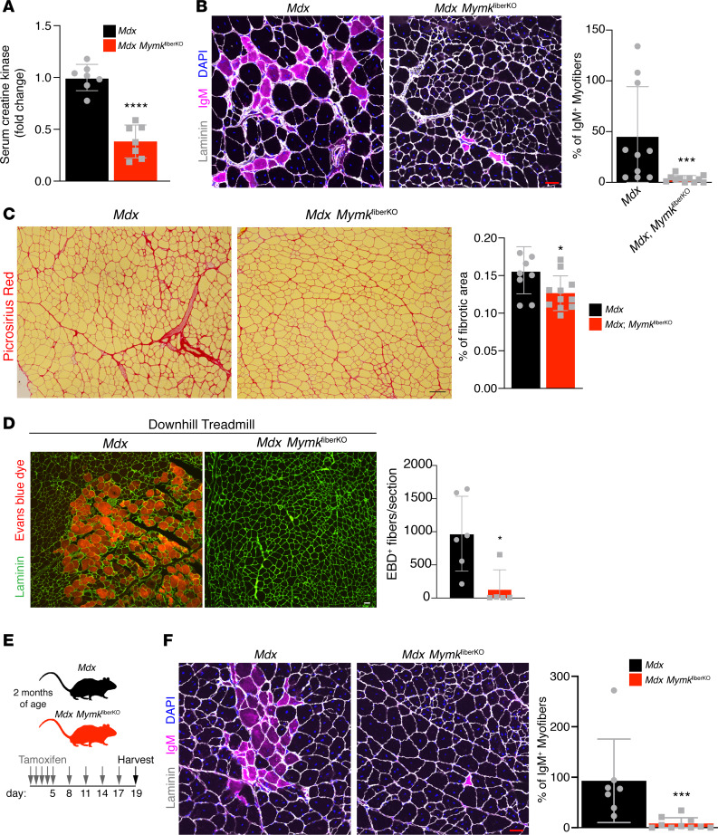
Figure 5. Deletion of Myomaker in myofibers leads to reduced muscle damage in mdx mice.
(A) Serum CK is reduced in mdx MymkfiberKO mice (n = 7). (B) IgM immunostaining of quadriceps (rectus femoris) sections shows reduction of damaged myofibers following deletion of myofiber-specific Myomaker expression (n = 8–11). (C) Picrosirius red stain of tibialis anterior (TA) muscle sections indicates less fibrosis in mdx MymkfiberKO mice (n = 8–11). (D) Rectus femoris muscle from mice treated with Evans blue dye (EBD) and subjected to a damage-inducing forced treadmill running protocol, with EBD+ myofibers quantified (n = 5–6). (E) Schematic of short-term mdx MymkfiberKO experiment showing continued tamoxifen administration for 2 weeks following initial 5-day induction. (F) Following short-term deletion of Myomaker in myofibers, IgM+ myofibers are reduced in rectus femoris muscle (n = 7–10). Statistical analyses and data presentation: (A and C) unpaired t test; *P < 0.05, and ****P < 0.0001; (B, D, and F) Mann-Whitney U test; *P < 0.05, and ***P < 0.001. Data are represented as mean ± SD. Scale bars: 50 μm (B, D, and F), 100 μm (C).

