
FIGURE 4.
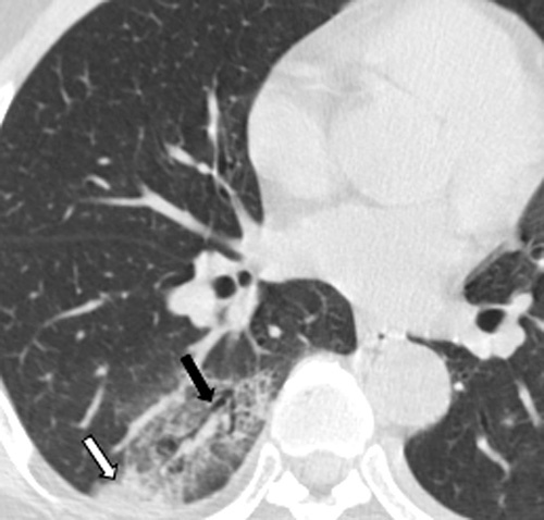
A 41-year-old man who presented with fever for 3 days. CT shows GGO with consolidation (white arrow) in the right lower lobe, accompanied by air bronchogram (black arrow).
Official websites use .gov
A
.gov website belongs to an official
government organization in the United States.
Secure .gov websites use HTTPS
A lock (
) or https:// means you've safely
connected to the .gov website. Share sensitive
information only on official, secure websites.
A 41-year-old man who presented with fever for 3 days. CT shows GGO with consolidation (white arrow) in the right lower lobe, accompanied by air bronchogram (black arrow).