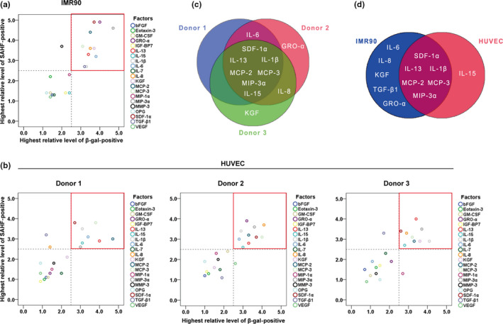
FIGURE 1.

Six pro‐inflammatory cytokines showed activities to induce cellular senescence. (a) Screening of cytokines on IMR90 cells, the values of “Highest relative level of SAHF‐ or β‐gal‐positive” were extracted from Table S5. The area circled by the red box in the upper right corner indicates the senescence inducers. (b) Screening of cytokines on primary cultured HUVECs from three donors, the values of “Highest relative level of SAHF‐ or β‐gal‐positive” were extracted from Table [Link], [Link], [Link]. (c) Venn diagram of three HUVEC strains. (d) Venn diagram of IMR90 cells and HUVECs
